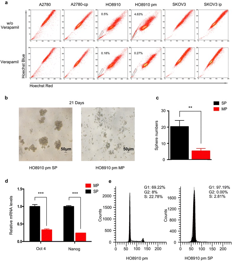
Fig. 1.

Separation of SP cells from the HO8910 pm cell line and verification of their stemness. a Detection of SP cells from high-grade serous ovarian cancer cells lines. Cells were stained with the dye Hoechst 33342 and analyzed by flow cytometry. The ratio of SP cells was highest in HO8910 pm cells. b The spheres that formed after 21 days of culturing in an ultra-low attachment plate were much larger of SP cells than MP cells. c More spheres formed of SP cells than MP cells. d The mRNA expression of Oct4 and Nanog was up-regulated in SP cells. e SP cells were arrested in G1 stage
