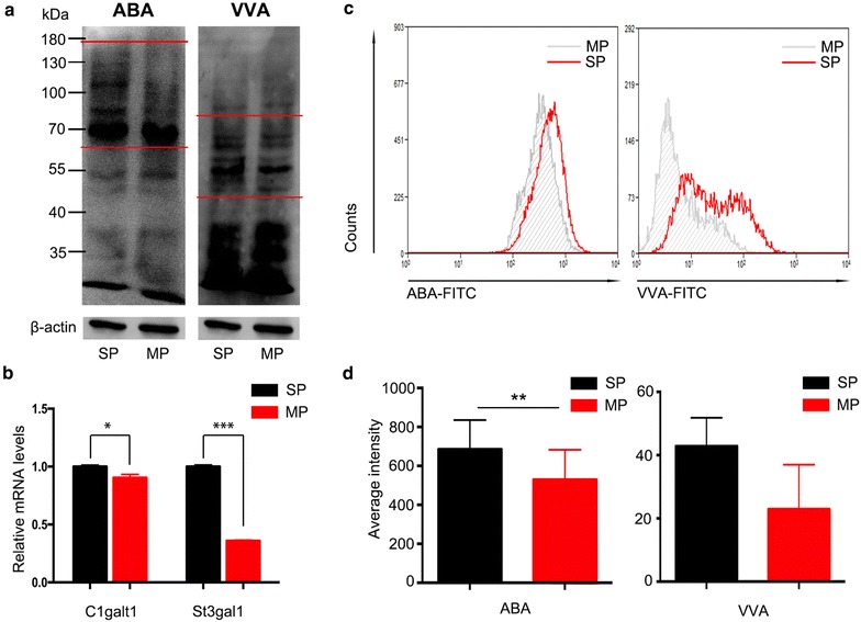
Fig. 5.

SP cellsexhibited higher intensity of ABA and VVA. a Lectin blots were used to detect the expression of the Tn (VVA, right), T and ST (ABA, left) antigens on SP cells and MP cells, β-actin was performed as control; b the mRNA expression levels of C1galt, which acts on Tn to form T antigen, and St3gal1, which forms ST antigen from T, were determined by real-time PCR. c, d The intensity of ABA and VVA for SP sphere cells after culturing for 5 weeks without serum was detected by flow cytometry and quantified
