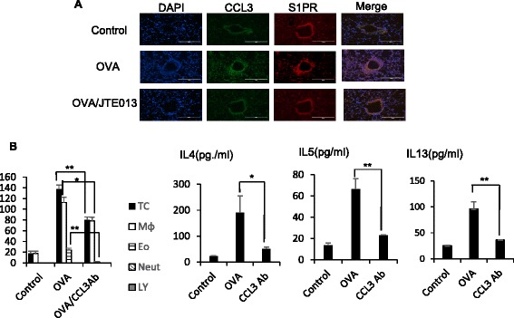
Fig. 3.

CCL3 and TIMP2 are S1P-dependent in vivo. Immunofluorescent microscopic images show OVA-treated lung sections stained with FITC-conjugated anti-CCL3 (green), and Alexa 594-conjugated anti-S1PR2 (red) antibodies. Co-localization of CCL3 and S1PR2 is seen in yellow (a). BAL Fluids were obtained from BALB/c mice treated with vehicle, OVA, or OVA/mCCL3 antibody (3 μg/cavity), and examined by Diff-Quick staining. The total cell counts and cell differentials in the BAL fluids are shown, and the concentrations of IL-4, IL-5, and IL-13 in the BAL fluids were measured by using ELISA kits (b). Data are expressed as the mean ± standard error (SE) of at least three independent experiments. *p < 0.05, **p < 0.01, ***p < 0.001
