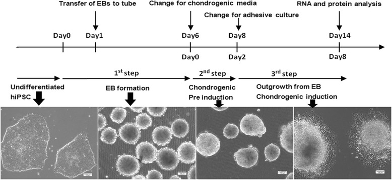
Fig. 1.

Schematic diagram of the culture protocol for chondrogenic differentiation of hiPSCs. The differentiation culture protocol consists of 3 steps: (1) Embryoid body (EB) formation in suspension culture dishes; (2) Pre induction of EB in the chondrogenic induction medium; (3) Cell outgrowth from EBs on gelatin-coated dishes in the chondrogenic induction medium. During differentiation, histological and gene expression analyses were performed at days 7 and 14. Scale bar: 100 μm
