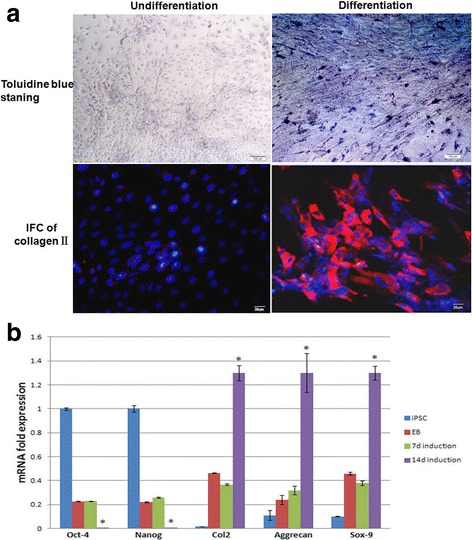
Fig. 2.

Examination of chondrogenic markers after differentiation. After EBs were differentiated for 2 weeks, staining with Toluidine blue revealed spherical cell morphology and interstitium, type II collagen was also detected after differentiation (a). The expression of chondrogenic differentiation markers was also analyzed by real-time PCR (b). Gene expression is normalized to that of beta–actin. Differentiated hiPSCs in monolayer culture exhibited high expression of COL2A1, ACAN and SOX9 compare with undifferentiated iPSCs. The expression was increased significantly at later stages (day 14). *Significant change relative to control (n = 4). Data are shown as the mean ± SD
