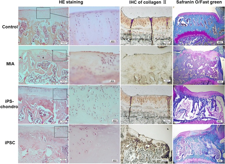
Fig. 6.

Histological analysis of knee sections. HE staining, Immunostaining of collagen II, Safranin O/Fast green staining were did to test the repairment of iPSCs. MIA-injected knee (MIA) showed obvious cartilage damage compare with normal knee (Control), while transplantation of iPS cells or iPS derived chondrocytes, the cartilage was repaired, and iPS derived chondrocytes (iPS-chondro) seems have stronger repair ability than iPS cells (iPSC)
