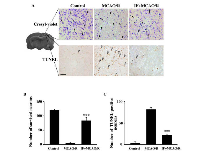
Figure 2.
IF rescues cortical neurons from MCAO/R-induced neuronal death via the attenuation of apoptosis. (A) Representative images of cresyl violet- and TUNEL-stained cortical regions in different groups at 72 h post-reperfusion (scale bar, 50 µm). Intact neurons are indicated by black arrows, dead or dying neurons with pyknotic nuclei are indicated by black arrowheads, and apoptotic neurons with TUNEL-positive nuclei are indicated by white arrowheads. (B) Quantitative analysis of intact cortical neurons (n=3/group; ***P<0.001 vs. MCAO/R). (C) Quantification of the number of TUNEL-positive neurons (n=3/group; ***P<0.001 vs. MCAO/R). Data are presented as mean ± standard error of the mean. IF, intermittent fasting; MCAO/R, middle cerebral artery occlusion and reperfusion; hippo, hippocampus; TUNEL, terminal deoxynucleotidyl transferase dUTP nick-end labeling.

