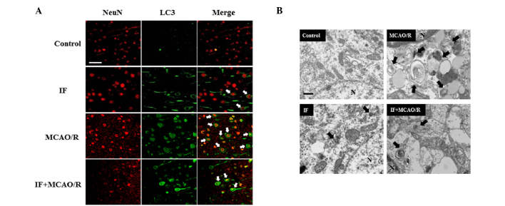
Figure 3.
IF inhibits the accumulation of APs in cortical neurons after MCAO/R. (A) Representative images showing immunofluorescent staining for LC3 (green, AP) and NeuN (red, neuronal nuclei) in the cortex at 24 h post-reperfusion with or without IF preconditioning. Neurons with autophagosomes are indicated by arrows in merged images (n=3/group; scale bar, 25 µm). (B) Representative electron microphotographs showing APs in the cortical penumbra of MCAO/R and IF + MCAO/R groups at 12 h after reperfusion (n=3/group; scale bar, 200 nm). Arrows indicate APs. IF, intermittent fasting; AP, autophagosomes; MCAO/R, middle cerebral artery occlusion and reperfusion; LC3, protein 1 lightchain 3.

