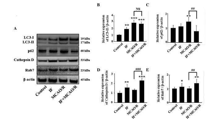
Figure 4.
IF enhances the autophagic flux in cortical neurons after MCAO/R. (A) Representative image of immunoblots for LC3, p62, cathepsin D, and Rab7 in cortical homogenates in different groups at 12 h post-MCAO/R. (B) Quantification of LC3-II protein level. These data were quantified as a proportion of LC3-I intensity (n=5/group; **P<0.01 and ***P<0.001 vs. control). (C-E) Quantification of p62, cathepsin D, and Rab7 protein levels, respectively. β-actin used as a loading control. (n=5/group; *P<0.05, **P<0.01 and ***P<0.001 vs. control; ##P<0.01 and ###P<0.001 vs. MCAO/R). Data are presented as the mean ± standard error of the mean. IF, intermittent fasting; MCAO/R, middle cerebral artery occlusion and reperfusion; LC3, protein light chain 3; NS, no statistical significance.

