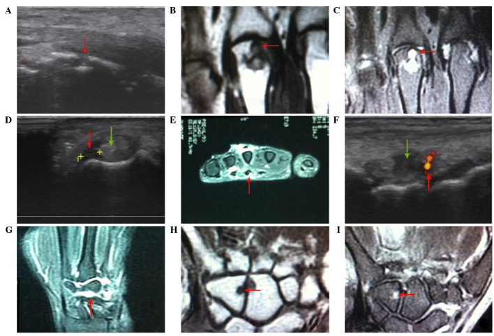
Figure 1.
Symptoms of abnormalities in rheumatoid arthritis with magnetic resonance imaging and high-frequency ultrasound. (A) Bone erosion (red arrow) indicated by ultrasound examination. (B) Bone erosion (red arrow) indicated by MRI featured by moderate signal intensities in the T1-weighted image. (C) Bone erosion (red arrow) indicated by MRI featured moderate signal intensities in the T2-weighted image. (D) Presence of tendonitis on the left hand by ultrasound examination, which demonstrated widening of the tendon (green arrow) and low intensity echoes around the tendon (red arrow). (E) Presence of tendonitis on the left hand indicated by MRI, which showed widening of tendon and high intensity T2-weighted image signals around the tendon (red arrow). (F) Presence of articular dropsy in the right wrist by ultrasound examination (red arrow). (G) Dropsy in the intercarpal joints and synovial proliferation indicated by MRI. (H) Presence of bone marrow edema by MRI, featured by low intensity T1-weighted image signals (red arrow). (I) Presence of bone marrow edema by MRI, featured by low intensity T2-weighted image signals (red arrow). MRI, magnetic resonance imaging.

