
Figure 3.
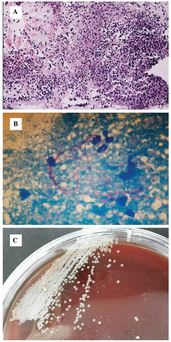
(A) Low power microscopy demonstrated a large amount of necrotic neutrophils, which indicated a purulent lesion. (B) Nocardia growth was confirmed by microscopy for a branching gram-positive rod, which was demonstrated to be positive for modified acid-fast stain. (C) Nocardia growth was observed by subculture, which demonstrated smooth and granular colonies.