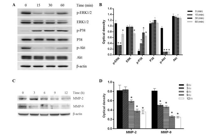
Figure 4.
Effect of emodin on the expression of proteins associated with tumor invasion. MHCC-97H cells were treated with 50 µmol/l emodin for the indicated times and the whole protein extracts were then analyzed by western blotting. (A) Representative western blot showing the protein expression of p-ERK, ERK, p-p38, p38, p-AKT and AKT. (B) Quantification of the relative protein expression levels were normalized against the value of β-actin protein expression. (C) Representative western blot showing the protein expression of MMP-2 and MMP-9. (D) Quantification of the relative protein expression levels normalized against β-actin protein expression. Data are presented as the mean ± standard deviation (n=3). *P<0.05 vs. the control group (0 min). p, phosphorylated; ERK1/2, extracellular signal regulated kinase; MMP, matrix metalloproteinase.

