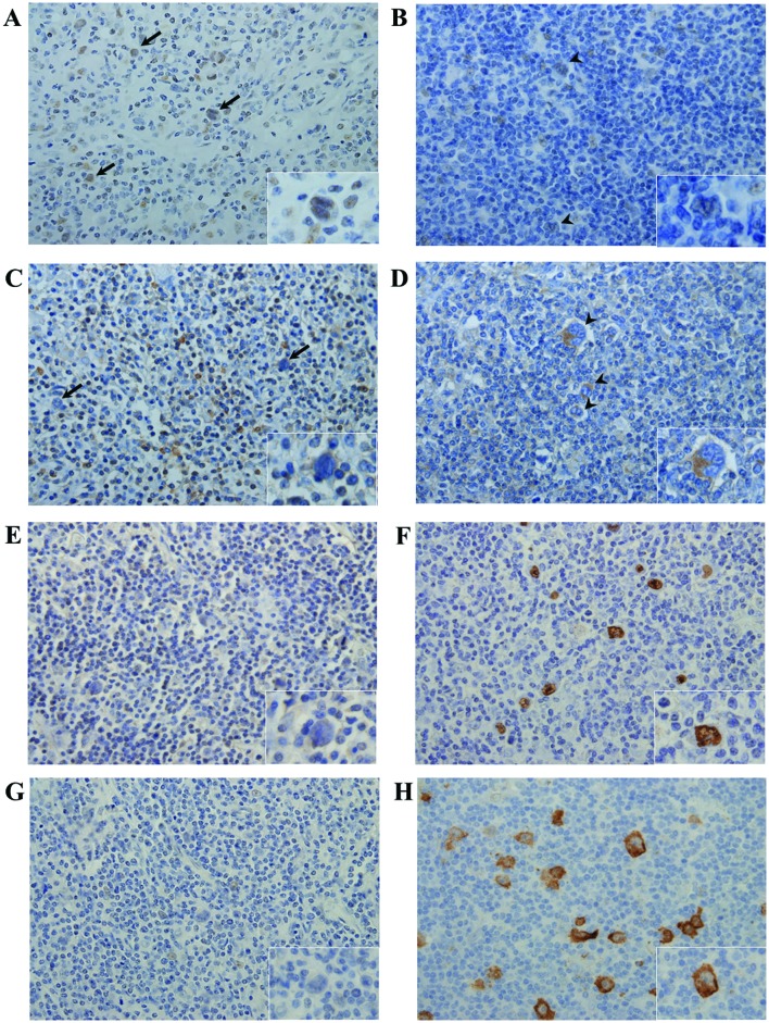
Figure 1.
FoxO3a, ID1 and LMP1 expression in HL. Typical cases [case 4 (A, C, E and G) and case 1 (B, D, F and H)] are shown. (A-D) Immunohistochemical detection of FoxO3a (A and B) and ID1 (C and D). Positive FoxO3a staining was detected in a limited number of tumor cells in case 4 (A) arrows and inset; magnification, ×400. In case 1, tumor cells (B) arrowheads and inset did not stain at all for FoxO3a, but certain non-tumor cells, such as macrophages and vascular endothelial cells, stained positive for FoxO3a (magnification, ×400). No ID1 signal was detected from tumor cells in case 4 (C) arrows and inset; magnification, ×400, whereas strong ID1 expression was evident in a limited number of tumor cells in case 1 (D) arrowheads and inset; magnification ×400. (E-H) EBER-1 in situ hybridization and immunohistochemical staining for the detection of LMP1. No EBER-1-positive/LMP1-expressing tumor cells were detected in case 4 (E and G), whereas tumor cells in case 1 stained positively for both EBER-1 and LMP1 (F and H). FoxO3a, Forkhead box O3a; ID1, inhibitor of DNA-binding protein 1; LMP1, latent membrane protein 1; EBER-1, Ebstein-Barr virus-encoded small RNA.

