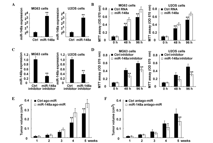
Figure 3.
Inhibition of miR-148a suppressed osteosarcoma growth both in vitro and in vivo. (A and B) MG63 and U2OS cells were transfected with control RNA or miR-148a mimics, as indicated. (A) miR-148a expression was examined using RT-qPCR. The data are the relative values of miR-148 expression normalized to the internal control of U6 expression. (B) Cell proliferation was measured using MTT assay at the indicated time points following transfection. (C and D) MG63 and U2OS cells were transfected with control RNA or miR-148a inhibitor, as indicated. (C) miR-148a expression was examined using RT-qPCR. The data are the relative values of miR-148 expression normalized to the internal control of U6 expression. (D) Cell proliferation was measured using MTT assay at the indicated time points following transfection. (E and F) MG63 cells were injected subcutaneously into the posterior flank of nude mice. Two weeks later, cholesterol-conjugated small RNA (E) ago-miR or (F) antago-miR of miR-148a were locally injected into the tumor mass, and tumor growth was measured. The growth curves obtained are indicated in the image. Data are presented as the mean ± standard deviation (n=4), or correspond to one representative experiment, whereby similar results were obtained in three independent experiments. *P<0.05; **P<0.01. miR, microRNA; Ctrl, control; RT-qPCR, reverse transcription-quantitative polymerase chain reaction; MTT, 3-(4,5-dimethylthiazol-2-yl)-2,5-diphenyltetrazolium bromide; OD, optical density; ago, agonist; antago, antagonist.

