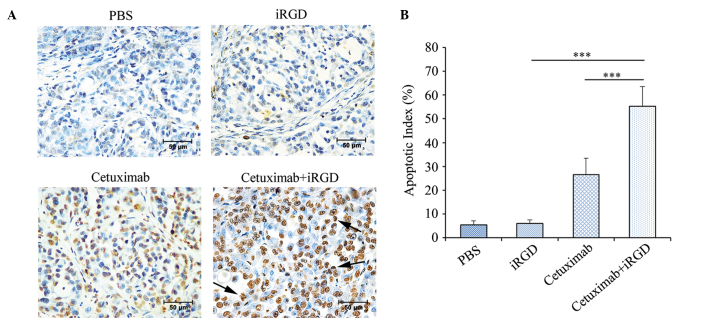
Figure 5.
Cancer cell apoptotic in the tumor co-administered with cetuximab and iRGD. (A) Apoptotic cells in the tumor tissues were detected by a TUNEL assay. TUNEL-positive nuclei are stained brown. TUNEL-negative nuclei are stained blue. The figures shown here are representative of the six tumors in each group. Arrows indicate the apoptotic bodies. Magnification, ×400; Scale bars=50 µm. (B) Quantitative analysis of the apoptosis index in each group. The percentage of TUNEL-positive cells counted from 100 randomly selected tumor cells per section. Five sections were counted per tumor. n=6; Error bars, mean ± standard deviation; ***P<0.001. PBS, phosphate-buffered saline; iRGD, internalizing RGD peptide; TUNEL, TdT-mediated dUTP nick end labeling kit.

