Figure 1.
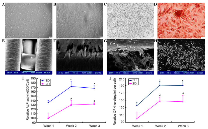
MSCs derived from acute myeloid leukemia patients were induced to form osteoblasts in vitro. (A) The morphology of the MSCs. (B) The morphology of the osteoblasts. (C) The cell identification was conducted by ALP staining. (D) The cells were identified by alizarin red staining. (E and F) The shape of the scaffolds and the osteoblasts seeded on the scaffolds. (G and H) The co-culture of the leukemia cells and osteoblasts in the 3D and 2D culture systems. (I and J) The levels of ALP and OPN increased, and the levels were higher in the scaffolds.*P<0.05 compared with the control group of the 3D system. #P<0.05 compared with the control group of the 2D system. MSC, mesenchymal stem cell; 3D, 3-dimensional; ALP, alkaline phosphatase; OPN, osteopontin; OD, optical density.

