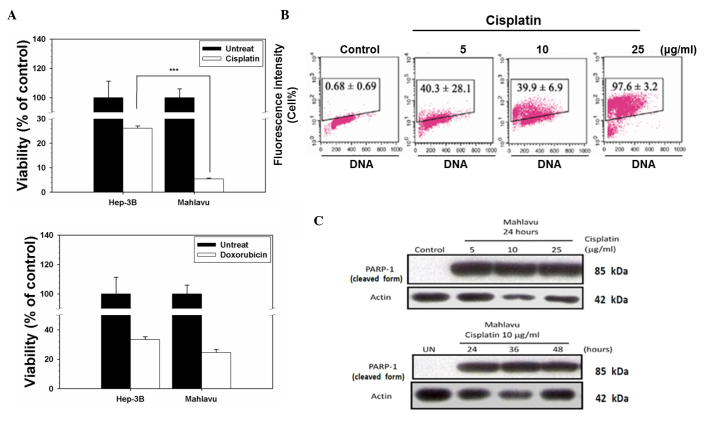
Figure 1.
Distinct disparity of sensitivity of hepatocellular carcinoma cells in response to chemotherapeutic drugs. (A) Mahlavu and Hep3B cells were treated with cisplatin or doxorubicin for 72 h. Cell viability was determined using a sulforhodamine B assay. The results are expressed as the mean ± standard deviation. Each point represents an average of ≥3 determinations. ***P<0.05 vs. control. (B) Evaluation of cisplatin-induced apoptosis in Mahlavu cells. Control (untreated) cells and cells treated with 5, 10 or 25 µg/ml cisplatin for 24 h were subjected to terminal deoxynucleotidyl transferase 2′-deoxyuridine 5′-triphosphate assay. The representative plots depict DNA content on the X axis and 5-bromo-2′-deoxyuridine-fluorescein isothiocyanate-labeled apoptotic DNA strand breaks on the Y axis. The data represent the percentage of apoptotic cells in the upper boxes. (C) The cleavage of PARP-1 (a 116-kDa nuclear enzyme downstream of caspase 3) into an 85-kDa fragment by cisplatin-treated cells provides further evidence of the occurrence of apoptosis through the activation of caspase 3. UN, untreated; PARP-1, poly(adenosine diphosphate-ribose) polymerase 1.

