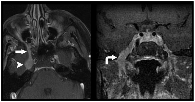
Figure 1.
Direct geographical invasion of primary head and neck cancer. 56-year old woman with no significant past medical history presented with 3 months right facial pain. Axial (left) and coronal (right) T1 weighted fat saturated post contrast MR imaging of the skull base demonstrates nodular enhancement of the right trigeminal nerve ganglion (arrow head) with mass like expansion of Meckle’s Cave. Linear thickening and enhancement is noted to extend through foramen rotundum (straight arrow) and foramen ovale (curved arrow). Tissue sampling of the lesion demonstrated Adenoid Cystic Carcinoma of the trigeminal nerve ganglion.

