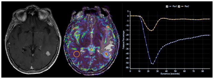
Figure 17.
DSC perfusion MR imaging characteristics of metastatic disease. Pre-operative MR imaging in a 63-year old woman with history of metastatic breast cancer demonstrates a contrast enhancing mass within the left temporal lobe. Cerebral blood volume map (center) and signal intensity/time curve (right) calculated from DSC perfusion MR imaging sequence (not shown) demonstrates elevated cerebral blood volume with reduced percentage of signal intensity recovery metrics (blue region of interest) when compared to contralateral normal appearing white matter (orange region of interest). Prior investigators have demonstrated that elevated cerebral blood volume within an enhancing mass is nonspecific for either high-grade glioma or metastatic disease. However, reduced percentage of signal intensity recovery values have been shown to be specific for the diagnosis of intra-parenchymal metastatic disease.

