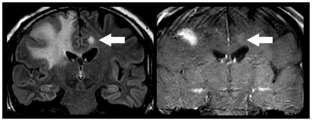
Figure 2.
Direct geographical invasion of Glioblastoma. 63 year old man with altered mental status presented for MR imaging. Coronal FLAIR (left) and T1 weighted post contrast (right) MR imaging of the brain demonstrates extensive FLAIR signal within the right cingulate, superior frontal, middle frontal, and inferior frontal gyri that extends across the corpus callosum to involve the contralateral cingulate gryus (arrow). Focal enhancement is noted within the ipsilateral mass and contralateral cingulate gyrus lesion. Subsequent resection of the right frontal mass demonstrated Glioblastoma. Multifocal glioma can occur at initial presentation with metastatic disease occurring via direct peri-neural infiltration.

