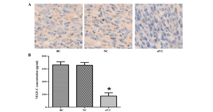
Figure 8.
VEGF-C protein expression in excised tumors of various groups. (A) Representative photographs of the tumor sections examined by immunohistochemical staining for VEGF-C (magnification, ×400). The assessment of VEGF-C expression was based on a cytoplasmic and intercellular substance-staining pattern. (B) Tumors were harvested and assayed for VEGF-C protein by an enzyme-linked immunosorbent assay. Each bar represents the mean ± standard deviation (n=6). *P<0.05 vs. BC group. VEGF-C, vascular endothelial growth factor C; RNAi, RNA interference; BC, blank control; NC, negative control small interfering RNA sequence transfection group; siV2, siV2 transfection group.

