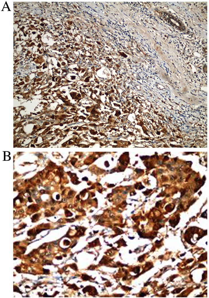
Figure 1.

Immunohistochemical staining of breast cancer tissue. (A) Strong nuclear and cytoplasmic HIF-1α positivity in the neoplastic breast carcinoma cells (left), and negative nuclear staining in the adjacent normal breast duct (upper right) (magnification, ×200). (B) High power view of strong nuclear and cytoplasmic HIF-1α expression in invasive breast carcinoma tissue (magnification, ×400). HIF-1α, hypoxia induciblefactor-1α.
