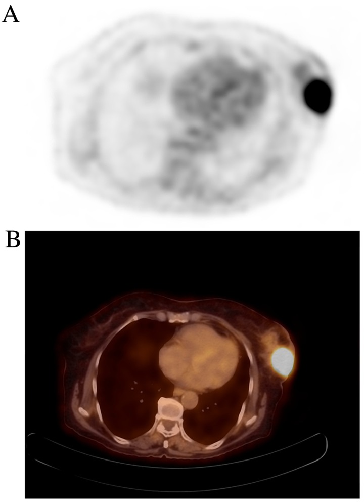
Figure 2.

(A) FDG PET and (B) hybrid PET/computed tomography axial images demonstrating a left breast cancer lesion with notably increased FDG uptake (maximum standardized uptake value, 11.9) in a 76-year-old woman with invasive breast carcinoma (no special type). Histopathological and immunohistochemical features of the tumor were as follows: pT2 (45 mm); N0; histologic grade 3; ER-; PgR-; HER-2 3+++; and Ki-67, 70%. FDG, 18F-fluorodeoxyglucose uptake; PET, positron emission tomography.
