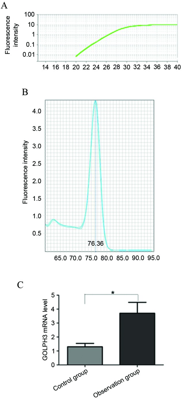
Figure 1.

Reverse transcription-quantitative PCR analysis of the GOLPH3 mRNA levels in the 2 groups. (A) PCR amplification curve. (B) The melting curve of PCR primers. (C) Comparison of GOLPH3 mRNA level in observation group and control group. *P<0.05. PCR, polymerase chain reaction; GOLPH3, Golgi phosphoprotein-3.
