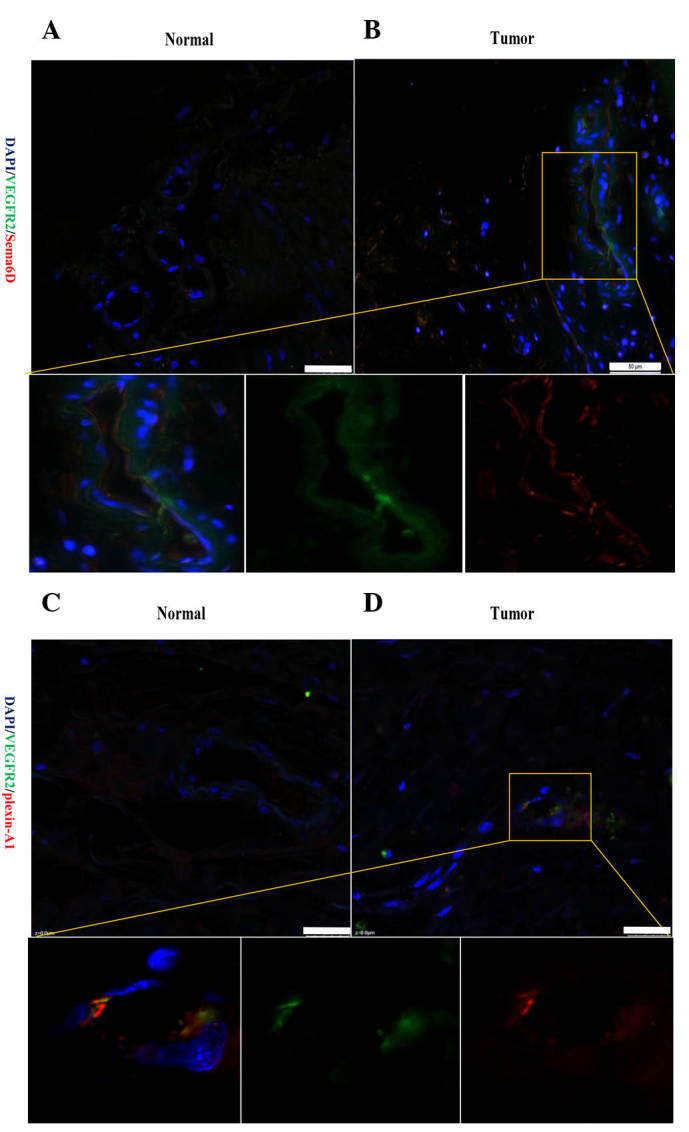
Figure 5.

Sema6D and plexin-A1 were expressed and positively correlated with VEGFR2 in vascular endothelial cells. (A and B) Representative fluorescence images showing the double-labelling of Sema6D and VEGFR2 in (A) peri-cancerous and (B) cancerous vessels. In the cancerous tissues, Sema6D was upregulated and present in the vascular epithelial cells. Furthermore, Sema6D expression was strongly correlated with VEGFR2 expression in vascular epithelial cells. (C and D) Compared with (C) normal vascular cells within gastric cancer, the double-labelling of cells for plexin-A1 and VEGFR2 could also be detected in (D) vascular epithelial cells. Bar =25 µm. Sema, semaphorin; VEGFR, vascular endothelial growth factor receptor; DAPI, 4′,6-diamidino-2-phenylindole.
