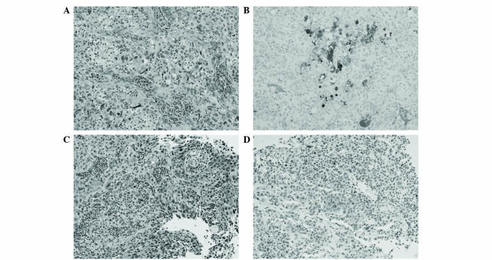
Figure 3.
(A and C) Hematoxylin & eosin staining of the tumor specimen showed poorly-differentiated urothelial carcinoma. Immunohistochemical staining results revealed (B) focally or diffusely positive reactivity for granulocyte colony-stimulating factor, and (D) scattered strong immunoreactivity for parathyroid hormone-related protein. Magnification, ×200.

