Figure 3.
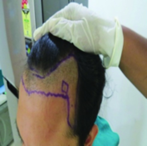
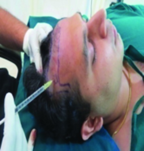
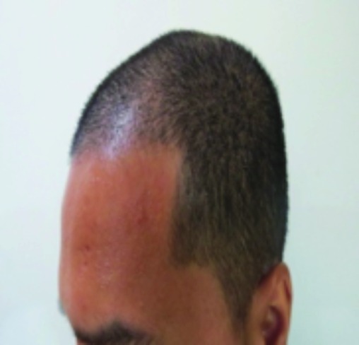
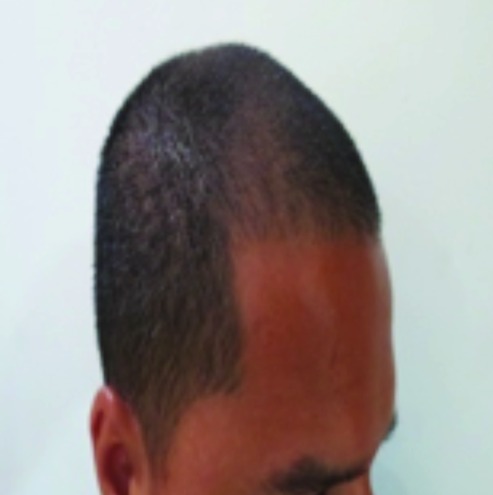
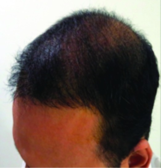
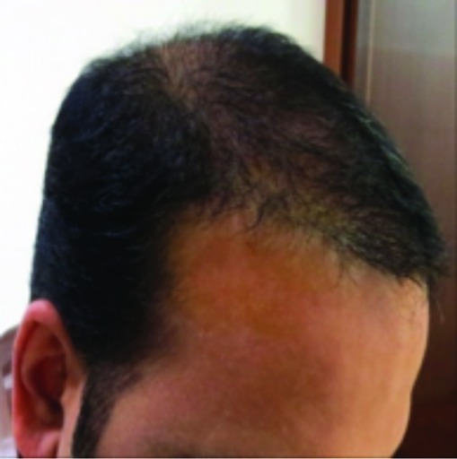
(A) Photographs showing the PRFM treatment being performed on the right side post-implantation and also the left side post-implantation. (B) Photographs taken after two months post-implantation and PRFM treatment. Right side of the scalp is PRFM treated post-implantation and left side of the scalp is the control: PRFM untreated post-implantation. (C) Photographs taken after six months post-implantation and PRFM treatment. Right side of the scalp is PRFM treated post-implantation and left side of the scalp is the control: PRFM untreated post-implantation.
