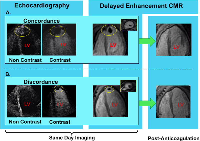
Figure 1. LV Thrombus Detection by Non-Contrast and Contrast Echo.

Representative examples of (1A) LV thrombus concordantly detected by non-contrast (left) and contrast (right) echo, and (1B) improved detection of LV thrombus via contrast echo. Images displayed in 4-chamber orientation (thrombus denoted by yellow circle).
In both examples, DE-CMR confirmed contrast echo results, as evidenced by thrombus-associated avascularity within the LV apex (images shown in 4-chamber [corresponding to echo], and short axis orientation [inset]). As shown on far right (green arrows), both cases also demonstrated resolution of DE-CMR-evidenced thrombus following treatment with warfarin-based anticoagulation.
