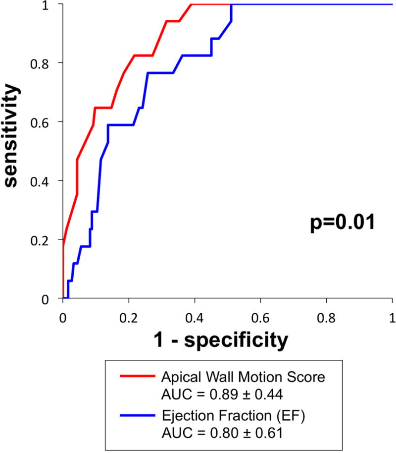
Figure 3. Receiver Operating Characteristics Curves.

Apical LV wall motion score via non-contrast echo (red) yielded improved diagnostic performance compared to EF (blue), as evidenced by higher area under the curve (p=0.01).

Apical LV wall motion score via non-contrast echo (red) yielded improved diagnostic performance compared to EF (blue), as evidenced by higher area under the curve (p=0.01).