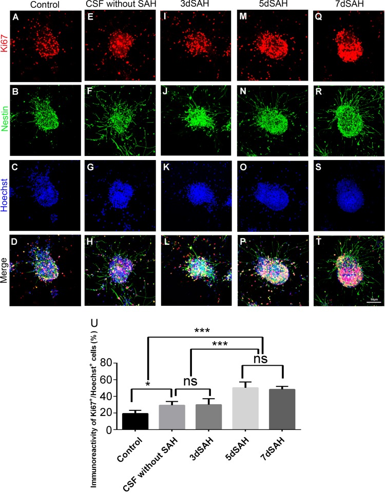
Fig 3. Treatment with post-SAH CSF promotes cell proliferation of cultured neurospheres.
Cells were immunostained with anti-nestin antibody (green: NSC), Ki67 antibody (red: proliferating cell) and Hoechst 33342 (blue: nucleus) to determine the level of cell proliferation of cultured neurospheres. (A)-(T): photomicrograph showing the distribution of Ki67+ (A, E, I, M, and Q) and nestin+ (B, F, J, N, and R) signals and as merged images (D, H, L, P, and T) in the neurospheres treated with and without CSF. Scale bar = 50 μm. (U): Percentage of proliferating cells (Ki67+ of Hoechst+ cells) in the neurospheres. Means ± SD, 12 neurospheres were counted for each condition. ns: non-significant; *, P < 0.05; ***, P < 0.001, one way ANOVA with Tukey's multiple comparisons test.

