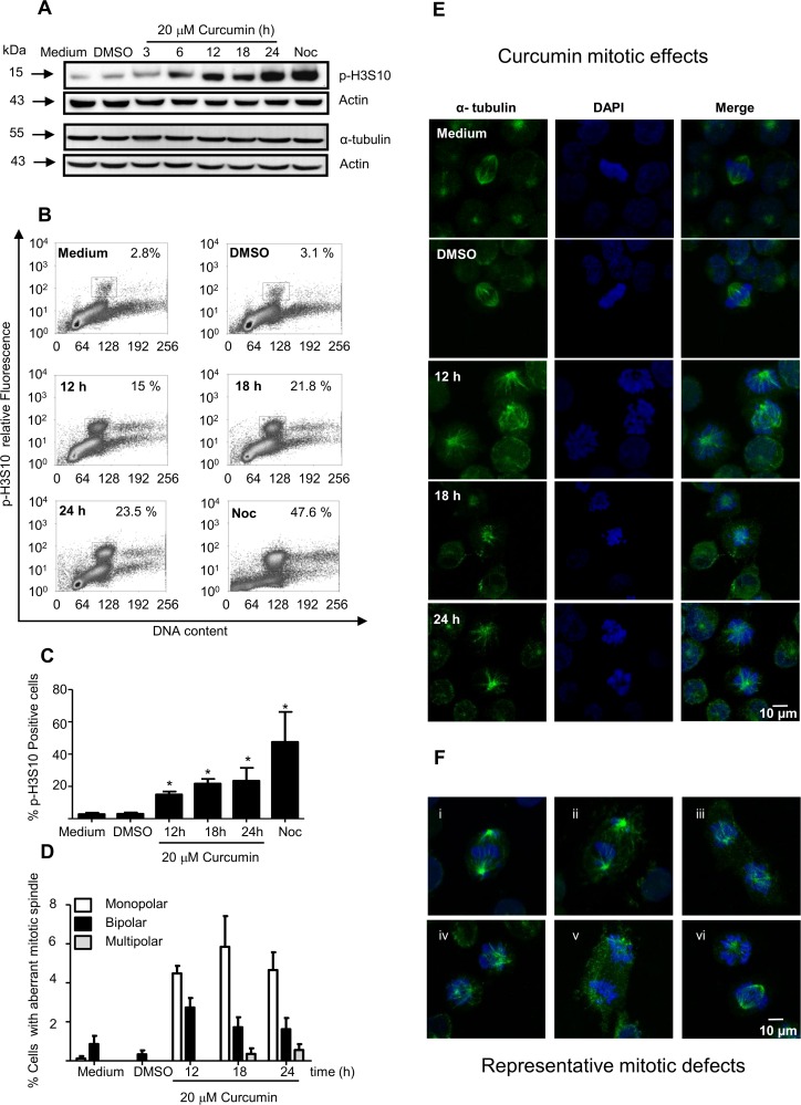
Fig 2. Mitotic spindles in K562 cells were altered by curcumin.
A) Expression levels of phosphorylsted-histone 3 in serine 10 (p-H3S10) were increased in K562 cells after curcumin treatment, as determined by western blot and B) FACS analysis. C) The relative numbers of p-H3S10-positive cells are presented as a histogram. K562 cells were stained with DAPI or immunostained using an antibody against α-tubulin (green fluorescence), and the cells with defects in the mitotic spindles were counted. D) The percentage of cells with monopolar (white bars), bipolar (black bars) and multipolar (gray bars) nuclei were determined in K562 cells. E) Representative fluorescence images of K562 cells bearing mitotic spindle alterations after 12 h, 18 h and 24 h of curcumin treatment are shown. F) Images of the most representative mitotic spindle alterations are shown. Actin was used as loading control.

