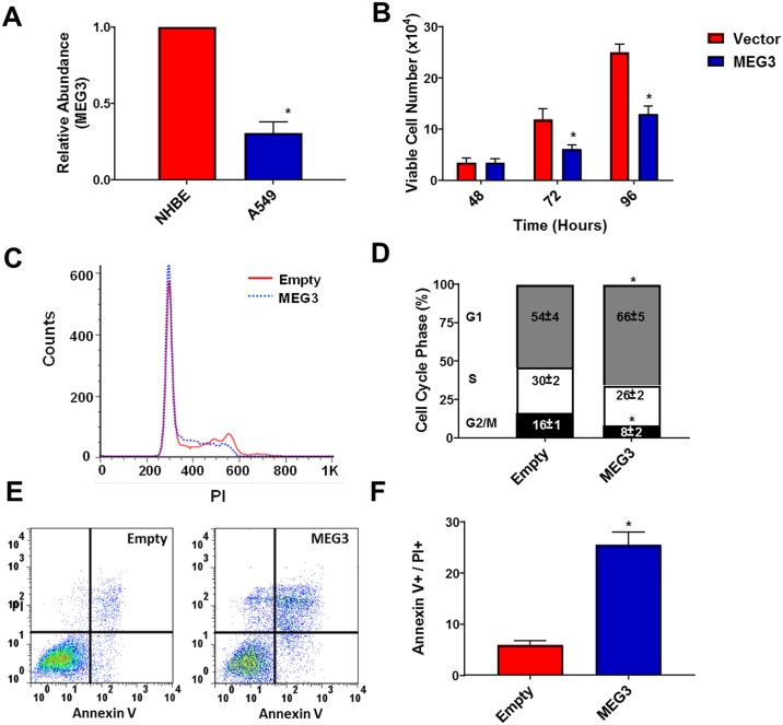
Fig 2. Inactivation of Rb leads to decreased expression of MEG3 and re-expression inhibits proliferation in A549 cells.
(A) Relative expression of MEG3 was determined by qPCR in NHBE and A549 (p16 deletion) lung cancer cells. (B) A549 cells were transfected with either a plasmid encoding human MEG3 or empty vector and viable cell number was determined at 48, 72 and 96 h Results are shown as mean ± S.D. for results from at least three independent experiments. (C&D) Distribution of the cells in the G1, S and G2/M phases of the cell cycle after 48 h by propidium iodide (PI) staining and flow cytometry. (E&F) Apoptotic rates were measured by the presence of PI and Annexin V positive cells after 48 h by flow cytometry. *p<0.05.

