
Figure 3.
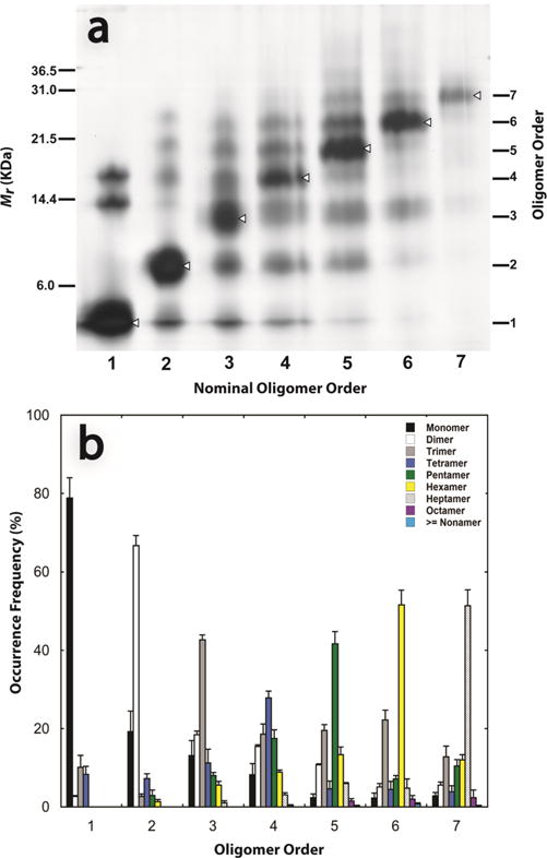
WT Aβ42 oligomer stability. Aβ42 was cross-linked and then electrophoresed in an SDS gel (see Materials and Methods). (a) Coomassie-stained oligomer bands were excised and re-electrophoresed, and the resulting bands visualized by silver staining. Each lane number represents the expected oligomer order. (b) ImageJ and MagicPlot were used to determine the occurrence frequencies of oligomers of each order. Data are representative of at least three independent experiments.