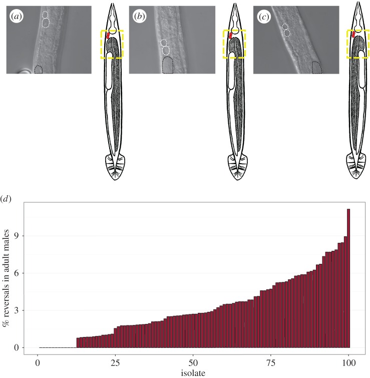
Figure 2.
The propensity for L/R gut/gonad reversals varies widely in males of 100 C. elegans isolates. (a–c) Examples of DIC images of ventral view showing the relative position of the gonad. Coelomocytes are marked with white-dashed lines and the top of the gonad arm is marked with black dashed lines. Dotted yellow box in the adjacent cartoon denotes the corresponding region in the photomicrograph in reference to the rest of the body. (a) Dextral N2 male, (b) dextral male from isolate JU778, (c) reversed JU778 male. (d) Percentage L/R gut/gonad reversals in adult males in the different natural isolates grown at 20°C. Each isolate is numbered 1–100 in rank order based on reversal frequency observed. Sample size per isolate ranges from 90 to 700 animals (a single replicate per isolate, except for isolates AB4, QX1211, N2 and CB4856, for which three replicates were performed and data were pooled when p > 0.05 using Fisher's exact test). See electronic supplementary material, table S1 for the key to the numbered isolate list and additional information.

