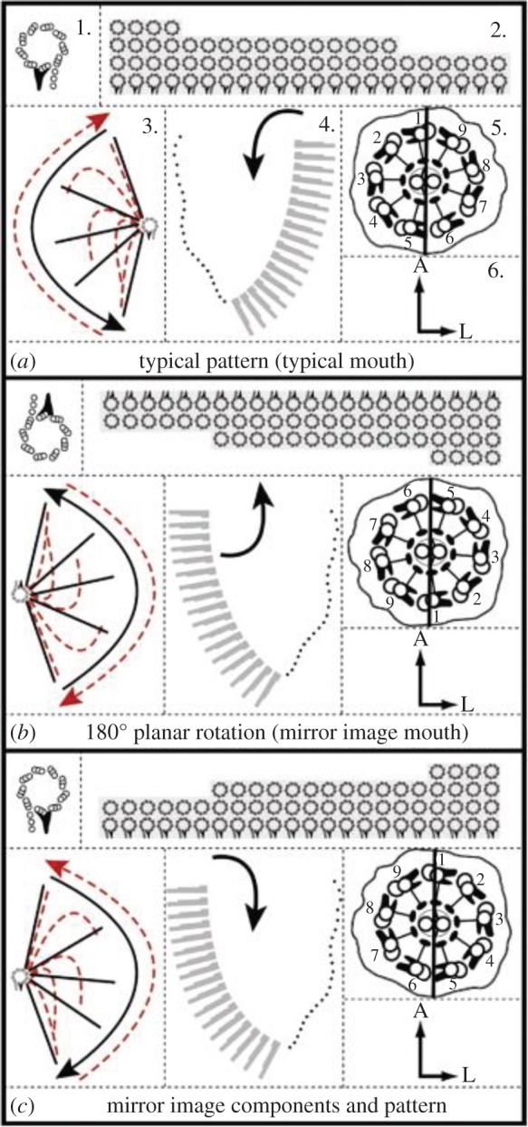
Figure 1.

Diagrams of Tetmemena oral apparatus showing basal body arrangement and ciliary numbering (a) wt left side of mirror image cell (b) mirror image right side, actual arrangement due to 180° rotation with no change of basal body chirality (c) hypothetical true mirror image of wt using reversed basal body chirality. (1) The basal body. Seen from outside the cell looking inward (tip to base). In (a) and (b), the basal body is drawn as observed with its true universal chirality. Looking tip to base, subfibres B and C of the triplet MT lie clockwise to subfibre A, the complete 13 protofilament MT. Attached to the basal body is the dense triangular basal foot that identifies the direction of the effective stroke of the cilia that grow on these basal bodies. This is reversed by the 180° in (b). In (c), the true mirror image requires a reversed basal body chirality where looking tip to base, subfibres B and C run anticlockwise from subfibre A, which is never observed in any cell. (2) The arrangement of the rows of basal bodies in the oral apparatus. The rotation in (b) requires a different order of morphogenesis. (3) The ciliary stroke. Solid lines indicate the effective stroke with direction of beat shown. Dashed lines indicate the recovery stroke. The 180° rotation reverses the direction of the effective stroke. (4) The direction of the current created by the cilia (black arrow). The current brings food into the wt oral apparatus and the cytostome. (5) The 9 + 2 ciliary cross-section with the doublets numbered. The chirality of the basal body is maintained. Looking tip to base, dynein arms occupy the free side of suberfibre A and run anticlockwise toward the adjacent subfibre B. The effective stroke is in the direction of doublets 5–6. (6) Positional coordinates of the oral apparatus with respect to the cell: A, anterior; L, left. Reproduced from Bell et al., [42] with permission. Courtesy A. Bell and Develop. Biol. (Online version in colour.)
