
Figure 7.
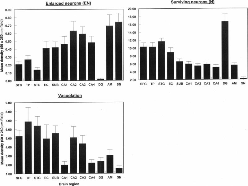
Densities of abnormally enlarged neurons (EN) and vacuoles (V) in various brain regions (SFG, Superior frontal cortex; TP, Temporal pole; STG, Superior temporal gyrus; EC, Entorhinal cortex; Sub, Subiculum; CA1-4, Cornu ammonis sectors of the hippocampus; DG, Dentate gyrus; AM, Basolateral amygdala; SN, Substantia nigra) in cases of chronic traumatic encephalopathy. Two-way analysis of variance (ANOVA): EN F = 6.81 (P < 0.001); V F = 3.81 (P < 0.001).