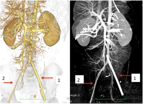
Fig. 4.

Three-dimensional computed angiogram performed after the primary surgery. Arrow 1: 12-Fr sheath in the left femoral artery. Arrow 2: right external iliac-femoral artery. Contralateral (right) external iliac-femoral artery appears thinner than the sheath placed in the left
