. 2016 Sep 30;18(3):306–314. doi: 10.7461/jcen.2016.18.3.306

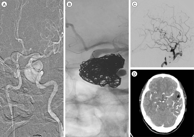
Fig. 4. Image taken during the second endovascular treatment. (A) The microcatheter can be seen entering the left distal internal carotid artery (ICA) via an A-Com channel, approaching from the right ICA, attempting to enter into the aneurysm through the gap between the stent and the ICA. (B) The aneurysm is packed using coils. However, the lower part of the aneurysm remained insufficiently packed. (C) In follow-up angiography, after coiling, reflux to the superior ophthalmic vein is found to be reduced. However, cortical venous reflux still exists. (D) On brain CT, low density and venous engorgement can be seen in the left temporal lobe. The asterisk indicates the infarct lesion. CT = computed tomography.

Fig. 4