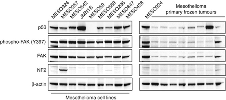
Figure 2.
Immunoblotting evaluation of the phosphorylation and expression of FAK and p53 in mesothelioma total cell lysates. The left panel shows mesothelioma cell lines and the right panel shows primary frozen tumours. Both western blots include one epithelial-type mesothelioma (MESO924) for comparison. Actin staining is a loading control.

