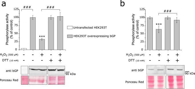
FIGURE 3.
bGP is inhibited in cells exposed to H2O2. a, HEK293T cells were transfected or not transfected with pCMV6-PYGB plasmid, and cells were exposed to 500 μm H2O2 for 20 min before being harvested. Cell lysis was then performed in the presence or absence of a reducing agent (10 mm DTT), and whole-cell extracts were assayed for endogenous glycogen phosphorylase activity. Data are expressed as the percentage of the control and represent mean values of three independent experiments ± S.D. ***, p < 0.001 when compared with positive control (upper panel); ###, p < 0.001 when two non-control groups are compared. Non-reduced and reduced whole-cell extracts were Western blotted and revealed for brain glycogen phosphorylase using an anti-bGP antibody. Ponceau red stains of the membranes are shown (lower panel). b, U87MG cells were exposed to 500 μm H2O2 for 20 min before being harvested. Cell lysis was then performed in the presence or absence of a reducing agent (10 mm DTT), and whole-cell extracts were assayed for endogenous glycogen phosphorylase activity. Data are expressed as the percentage of the control and represent mean values of three independent experiments ± S.D. ***, p < 0.001 when compared with positive control (upper panel); ###, p < 0.001 when two non-control groups are compared. Western blotting analysis of bGP from cells was revealed for brain glycogen phosphorylase using anti-bGP antibodies. Ponceau red stains of the membranes are shown (lower panel).

