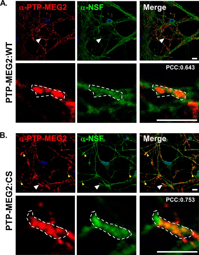
FIGURE 2.
Localization of PTP-MEG2 and NSF in the neurites of cortical neurons. Cortical neurons transduced with the lentiviruses expressing PTP-MEG2 (A) and PTP-MEG2:C515S (B), respectively, were immunostained with the antibodies for PTP-MEG2 and NSF, as indicated in red and green, respectively. Shown are typical confocal images of PTP-MEG2, PTP-MEG2:C515S, and NSF distribution in these neurons. DAPI staining indicates the location of nuclei (blue). Small yellow arrowheads indicate co-localization of PTP-MEG2:C515S and NSF in the neuronal puncta, and large arrowheads indicate regions enlarged with higher magnification. Low magnification scale bar = 10 μm; high magnification scale bar = 5 μm. Dotted areas indicate regions of interest for calculation of Pearson's correlation coefficient (PCC) of red and green fluorescence in a single Z section with compensation and subtraction of background fluorescence in both channels using Volocity software (PerkinElmer Life Sciences).

