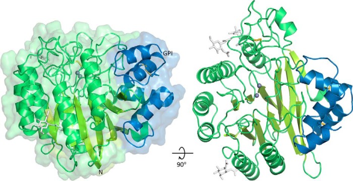
FIGURE 1.
Crystal structure of SMPDL3B. The catalytic domain is shown in green, and the C-terminal subdomain is in blue. Zinc ions are represented by gray spheres. N-Linked glycans (white sticks) are only partially displayed for clarity. Disulfide bonds are shown as yellow sticks. The N terminus and the putative C-terminal ω-site of GPI anchor attachment are indicated.

