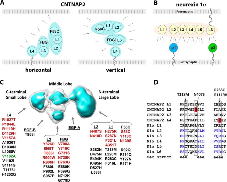
FIGURE 10.
Structural architecture and functional relationships of CNTNAP2. A, possible orientations of the CNTNAP2 ectodomain in the cleft of synaptic and axo-glial contacts. B, a horizontal orientation of the presynaptic neurexin 1α ectodomain at synaptic clefts promotes binding of protein partners tethered to the postsynaptic membrane (p1 and p2). C, location of amino acid substitutions in CNTNAP2 found in patient (red) and control (black) groups or both (green) as described in the “Results” section. D, sequence alignment of LNS domains from CNTNAP2 and neurexin 1α. Secondary structure prediction is shown (e is β-strand). Mutations discussed are indicated.

