FIGURE 8.
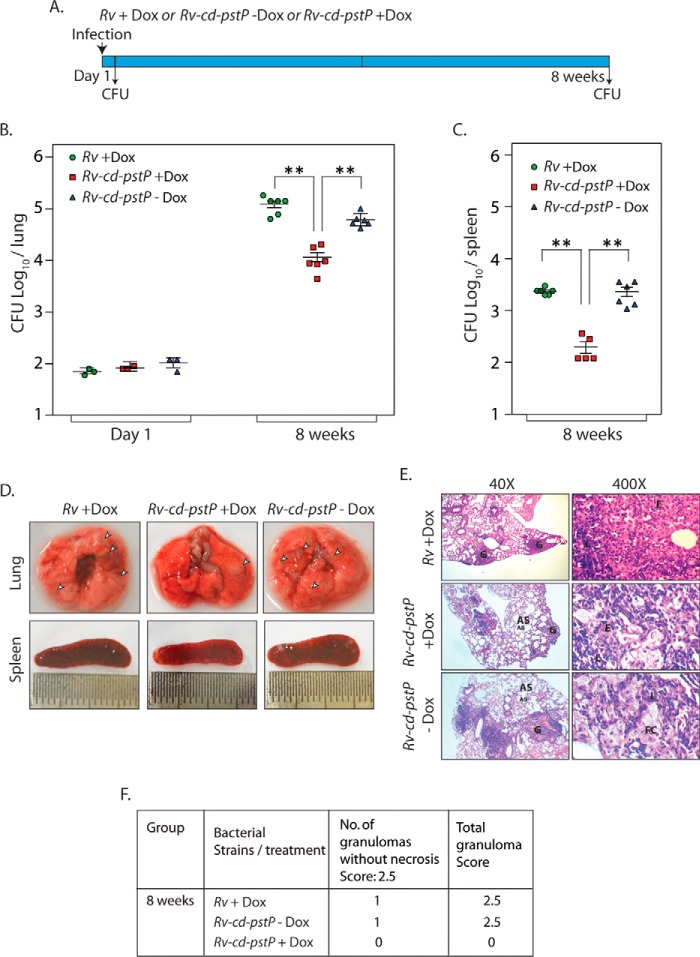
Depletion of PstP in M. tuberculosis negatively impacts its survival in mice. A, schematic representation of the mouse infection experiment. B and C, BALB/c mice were infected with Rv (9 mice) and Rv-cd-PstP (18 mice) as described previously (50). Rv-cd-PstP-infected mice were divided into two groups, and one group was provided Dox (1 mg/kg) in their drinking water. Rv-infected mice (9 mice) were also provided Dox in their drinking water. The water was changed every third day. Three mice from each group were sacrificed 1 day post-infection, and lung homogenates were plated in duplicates to determine mycobacterial implantation efficiency. The six remaining mice per group were sacrificed 56 days post-infection, and lung and spleen homogenates were similarly plated. B, the mean log10 cfu obtained for lung homogenates from Rv, Rv-cd-PstP +Dox, and Rv-cd-PstP −Dox at day 1 and day 56 were 5.1, 4.0, and 4.8 cfu, respectively. C, the mean log10 cfu obtained for spleen homogenates from Rv +Dox, Rv-cd-PstP +Dox, and Rv-cd-PstP at day 56 were 3.3, 2.1, and 3.3, respectively. Error bars, S.E. **, p < 0.01, two-tailed nonparametric t test. D, gross lung and spleen pathology of Rv +Dox-, Rv-cd-PstP +Dox-, and Rv-cd-PstP-infected mice 8 weeks post-infection. Prominent tubercles are indicated by white arrowheads. E, ×40 and ×400 images of H&E-stained lung sections of the infected mice. G, granuloma; FC, foamy cells; AS, alveolar space; E, epithelioid cells. F, granuloma score of the histopathology sections shown in E.

