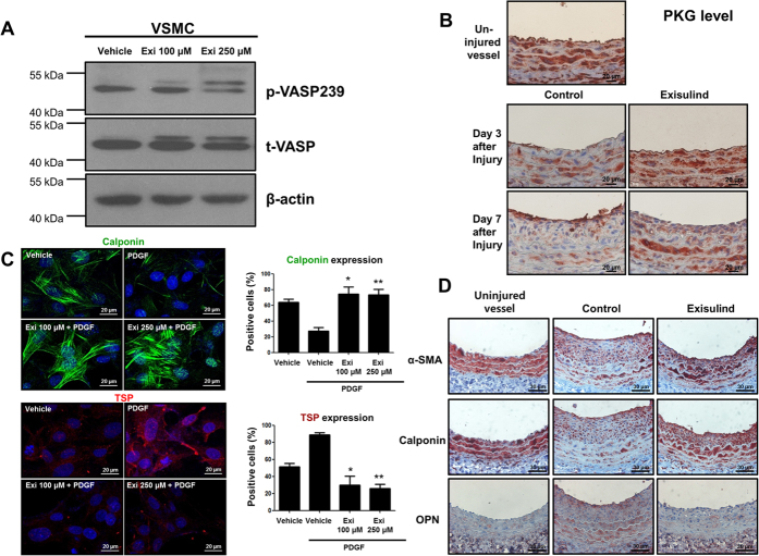
Figure 3. Effects of Exisulind on PKG activity and VSMC phenotype.
(A) Western blot analysis shows that Exisulind treatment increased VASP phosphorylation (a substrate of PKG) in a dose-dependent manner. p-VASP 239 = phospho-VASP at serine239, t-VASP = total VASP, Exi = Exisulind. (B) Immunohistochemical staining for PKG in rat carotid arteries. After balloon injury, PKG level was decreased. Compared with vehicle-treated group, Exisulind treatment reversed the decreased level of PKG induced by vascular injury. Scale bar = 20 μm. (C) Immunofluorescence staining for calponin and thrombospondin. PDGF treatment reduced the level of calponin (a marker for the differentiated contractile form of VSMC) and elevated the level of thrombospondin (a marker of synthetic from). Exisulind treatment reversed the effect of PDGF. Scale bar = 20 μm. Exi = Exisulind. *P < 0.01 vs. PDGF group, **P < 0.01 vs. PDGF group in terms of calponin level; *P < 0.01 vs. PDGF group, **P < 0.01 vs. PDGF group in terms of thrombospondin level. The data are presented as mean + SEM of four to five independent experiments. (D) Immunohistochemical staining for calponin, α-SMA, and osteopontin. The uninjured vessels showed the high level of α-SMA and calponin, but low level of osteopontin. The arterial wall at 2 weeks after injury showed that Exisulind treatment increased the expression of α-SMA and calponin (a contractile form marker) and decreased the level of osteopontin (a synthetic form marker). Scale bar = 20 μm. OPN = osteopontin.

