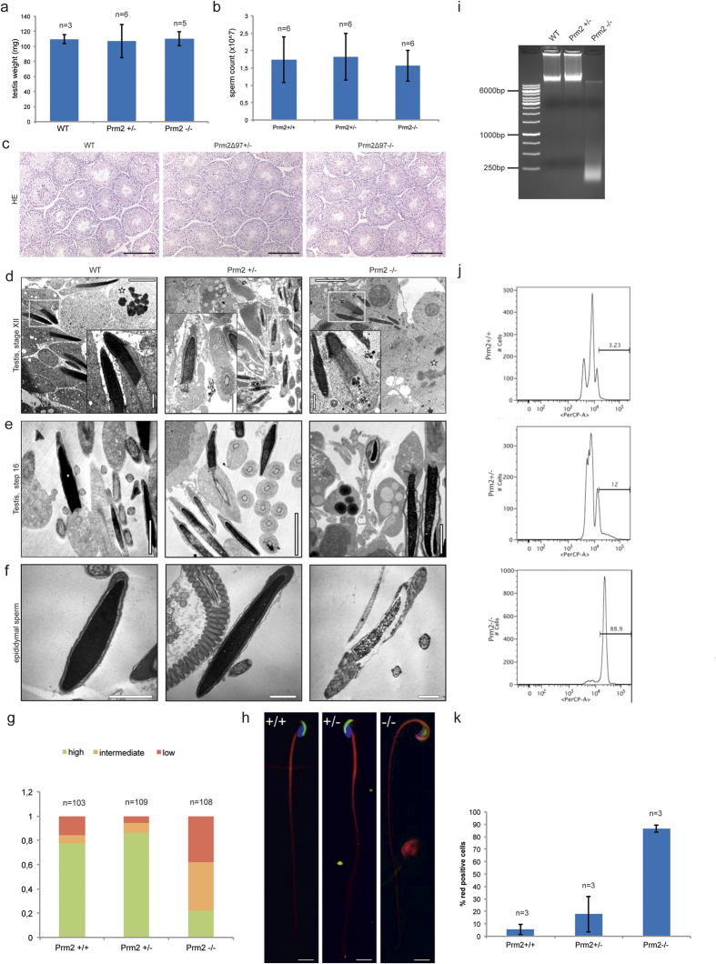
Figure 3. Morphological testes and sperm analysis.
(a) Testes weight. (b) Epididymal sperm count of Prm2+/+, Prm2+/−, and Prm2−/− mice. (c) HE staining of testicular sections from wildtype, Prm2+/− and Prm2−/− mice. Scale bar = 200 μm. (d) Transmission electron micrographs (TEM) of stage XII seminiferous epithelium as indicated by meiotic spermatocytes (stars) showing step 12 elongating spermatids. Scale bar = 5 μm. Inset: enlargement of rectangle, scale bar = 1 μm. Note homogeneously condensed chromatin in wildtype in contrast to heterogeneously condensed fine-grained chromatin in the Prm2-deficient mutant, whereas formation of the acrosome and manchette as well as head shape did not differ. (e) TEMs of step 16 elongated spermatids prior to sperm release. Scale bar = 1 μm. (f) TEMs of epididymal sperm. Note nuclear matrix alteration, detachment of acrosome (arrow) and attachment of the flagellum to the head (arrowhead) in knockout sperm. Scale bar = 1 μm. (g) Quantification of epididymal sperm chromatin integrity. (h) Prm2−/− but not Prm2+/− sperm showed morphological defects in the acrosome and head-tail conjunction. The acrosome was labeled using PNA-FITC (green), DNA using DAPI (blue), and the flagellum using MitoTracker (red). Scale bar = 10 μm. (i) Agarose gel electrophoresis of sperm genomic DNA. (j,k) Sperm damage analysis referring to SCSA.

