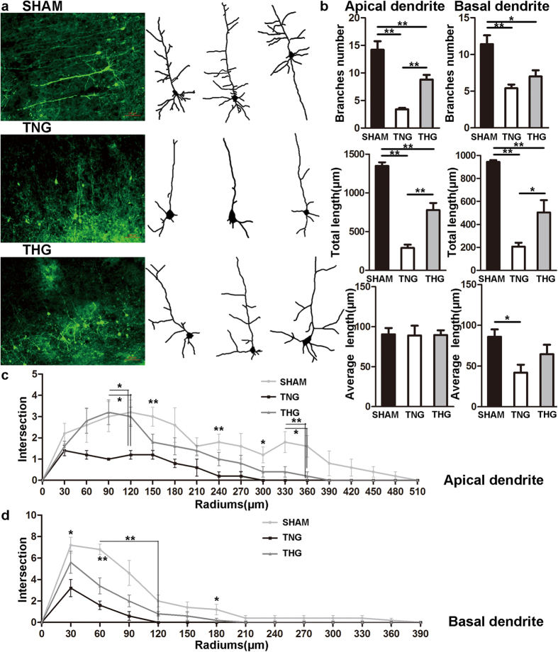
Figure 1. Posttraumatic hypothermia prevented dendrite degeneration 1 day after severe TBI.
(a) Image data were collected using fluorescence microscope. Left panel: Cortical pyramidal neurons within 2 mm of the lesion cavities of the three groups. Right panel: Reconstruction of the cortical pyramidal neurons in the three groups. (b) Branches number, total length and average length were compared across the three groups. Compared with the TNG, the THG showed increased branch number and total length in apical dendrites as well as increased total length in basal dendrites. (c,d) A Sholl analysis was used to assess the observed changes in dendrite complexity. Compared with the TNG, the THG had more intersections 90–120 μm from the neuron soma in the apical dendrites. The data are represented as mean ± SEMs and analysed using one-way analysis of variance (ANOVA) followed by Tukey’s post hoc test, n = 10, *p < 0.05, **p < 0.01, THG or sham versus TNG. TNG, the traumatic brain injury with normothermia group; THG, the traumatic brain injury with hypothermia group.

