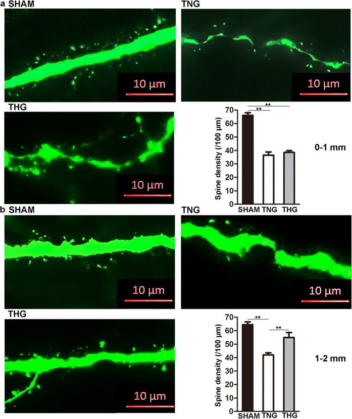
Figure 3. Posttraumatic hypothermia prevented spine loss 1 day after severe TBI.
(a) Representative images and quantitative analyses of spines (right bottom panel) in the three groups within 0–1 mm from the edge of the lesion cavity. (b) Representative images and quantitative analyses of the spines (right bottom panel) of three groups within 1–2 mm from the edge of the lesion cavity. High-resolution images were captured using fluorescence microscopy at a magnification of 630×. The data are represented as the means ± SEMs and analysed using one-way analysis of variance (ANOVA) followed by Tukey’s post hoc test, n = 10, **p < 0.01, THG or sham versus TNG. TNG, the traumatic brain injury with normothermia group; THG, the traumatic brain injury with hypothermia group.

