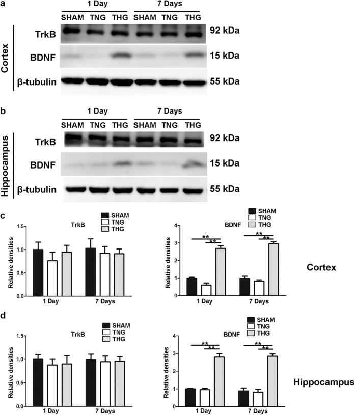
Figure 7. Temporal profile of BDNF and TrkB protein expression levels after severe TBI.
(a) Western blot analysis was performed to evaluate the BDNF and TrkB expression levels in the cortex 1 and 7 days after injury. (b) Western blot analysis was performed to evaluate the BDNF and TrkB expression levels in the hippocampus 1 and 7 days after injury. (c,d) Quantitative analyses of the BDNF and TrkB expression levels in the cortex and hippocampus were performed. The results of the quantitative analyses were normalized to β-tubulin levels. The data are represented as the means ± SEM and analysed using a one-way analysis of variance (ANOVA) followed by Tukey’s post hoc test, n = 10, **p < 0.01, THG or sham vs. TNG. TNG, the traumatic brain injury with normothermia group; THG, the traumatic brain injury with hypothermia group.

