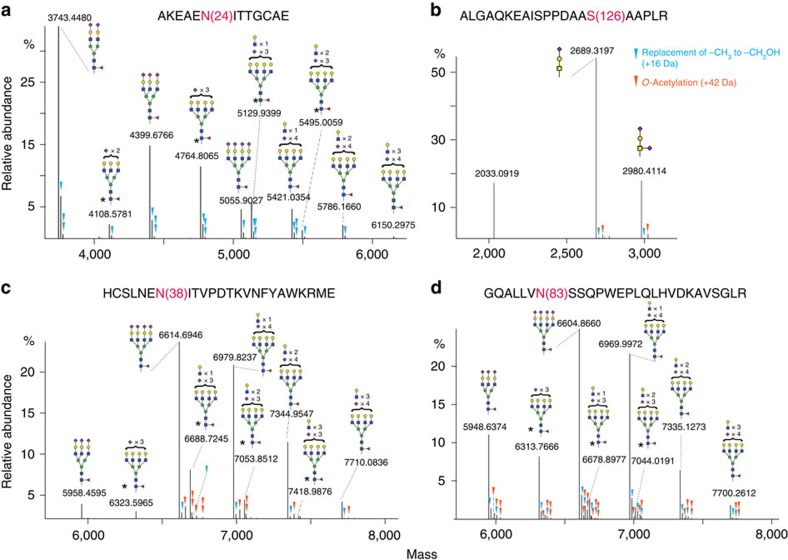
Figure 3. Middle-down proteomic analysis of rhEPO.
PTM profiling of site-specific glycoforms of rhEPO, covering the three N-glycosylation sites N24 (a), N38 (c), N83 (d) and the O-glycosylation site S126 (b). Of note, all the presented triantennary N-glycans may contain two types of structural isomers: α1–3 triantennary (shown in the figure), and α1–6 triantennary structures. * indicates the possibilities of a second co-existing glycan structure in addition to the one shown in the figure. For example, a triantennary structure with two sialic acids can also be a diantennary structure with an extra Galβ-1,4GlcNAc unit; a tetra-antennary structure with three sialic acids can also be a triantennary structure with an extra Galβ-1,4GlcNAc unit. The triangles indicate additional modification of sialic acids by either O-acetylation (in orange) or the replacement of −CH3 by −CH2OH (in blue).

